Under the hood
Modeled, not mimicked.
Nothing on screen is an animation, a lookup table, or a recorded waveform. Every
electrogram, every surface lead, and every response to pacing is computed, beat by
beat, from the same simulated tissue — so when the physiology changes, everything
downstream changes with it.
That is the difference between a simulator and a picture of one.
- Atria
- Reaction–diffusion across a biatrial mesh with anisotropic conduction, ionic membrane models, and a two-layer wall where fibrillation needs one.
- Ventricles
- Volumetric, tetrahedral myocardium with a fiber field and endocardial-to-epicardial heterogeneity, running a published human ventricular action-potential model.
- AV node
- Dual pathways with decremental conduction, so nodal reentry, Wenckebach, and block arise from the node rather than a rule.
- His–Purkinje
- A Purkinje network with loops that conducts in both directions and takes part in ectopy and arrhythmia.
- Electrograms
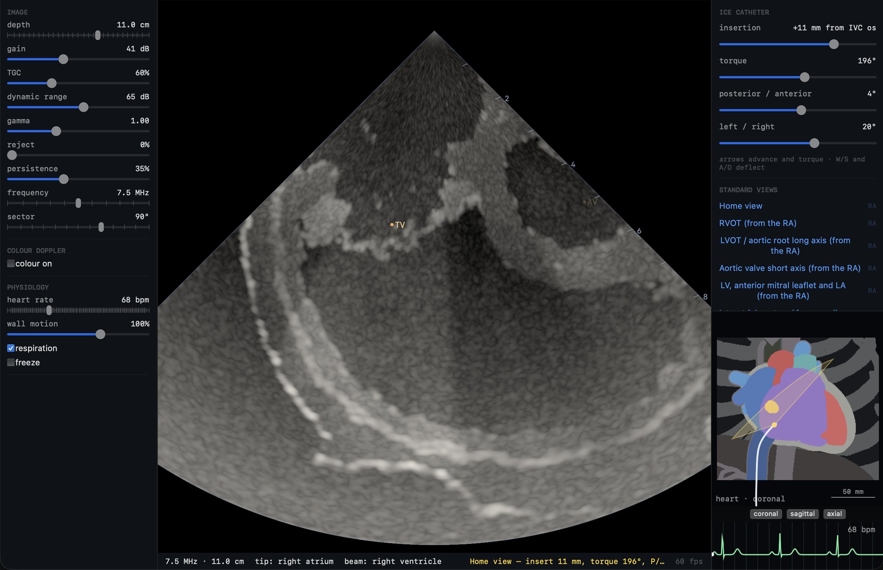
- Unipolar potentials from the tissue’s own current sources; bipoles are their difference, filtered as a recording system filters them.
- 12-lead ECG
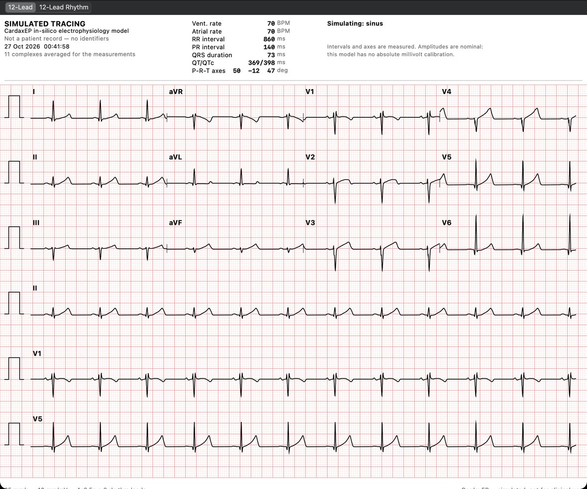
- The same sources summed at the body surface: axis, intervals, R-wave progression, and PVC morphology follow from the activation sequence.
- PVC origins
- Twenty-five ventricular origins placed by anatomy, at a chosen depth through the wall — the 12-lead and the activation map follow from where the beat begins.
- Mapping
- Activation and voltage maps built from catheter electrograms, beat by beat, with the beat gating a clinical system uses.